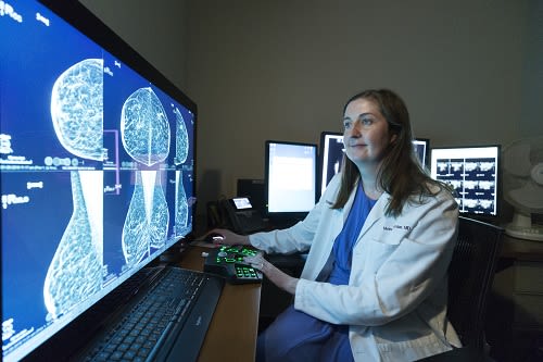
By Moira O’Riordan, Director, Women’s Imaging and Co-Director, Stamford Health Breast Center
Whether you’re about to turn 40 and scheduled your first mammogram, or you’ve had a mammogram and you were told you might need a breast ultrasound, the following Q&A’s with Moira O’Riordan, Director, Women’s Imaging and Co-Director, Stamford Health Breast Center, aims to answer all questions you might have.
There are many reasons for a breast ultrasound, but the most simple is:
Learn more about dense breasts and why a screening ultrasound is effective for examining dense breast tissue.
Remember to book your annual mammogram at Stamford Health’s Breast Center. You can choose from one of four convenient locations.
Book Your 3D Mammogram Today
For more information and to schedule an appointment, click below.
Book Appointment
Results given during business hours (8AM to 4PM, M-F.) For after-hours screening, results the next business day.
Book Appointment
Results given during business hours (8AM to 4PM, M-F.) For after-hours screening, results the next business day.
Whether you’re about to turn 40 and scheduled your first mammogram, or you’ve had a mammogram and you were told you might need a breast ultrasound, the following Q&A’s with Moira O’Riordan, Director, Women’s Imaging and Co-Director, Stamford Health Breast Center, aims to answer all questions you might have.
Why would I need a screening breast ultrasound after a mammogram?
Breast ultrasounds, if needed, are typically done with a mammogram. When used together, they help breast radiologists (also known as your doctor) get a better read for women with dense breasts.There are many reasons for a breast ultrasound, but the most simple is:
You have dense breast tissue.
Size and shape aren’t the only factors when it comes to breasts. Dense breast tissue can sometimes make it difficult to see small masses that may be “hiding.” That’s why ultrasound allows us to get a more complete picture of your breasts, along with a mammogram.Learn more about dense breasts and why a screening ultrasound is effective for examining dense breast tissue.
What happens during a screening breast ultrasound?
A technician will bring you to another room where you’ll lie down on a table. He or she will apply warm gel to your breast and then move a wand around your breast to take images of your tissue. If something is found, the technician will measure those findings for continued monitoring and establish a baseline together with your doctor.What can you find during a screening breast ultrasound?
There are a number of factors doctors look for when reading your ultrasound:- Normal tissue – Often feels lumpy in texture and is different for every women.
- Cysts – Sacs filled with fluid which are usually not cancerous (benign)
- Fibroadenomas — Solid, non-cancerous breast lumps that happen most often in women between 15 and 35 years old.
- Mass – Abnormal tissue that can be cancerous or non-cancerous.
How often will you need an ultrasound?
Depending on your breast density, your doctor may encourage you to get an ultrasound every year along with your mammogram. Sometimes, you may need to have an ultrasound every six months to watch your breast tissue more frequently, and monitor for any changes to size and shape. This process generally takes between 20 and 30 minutes.What happens once your ultrasound is complete?
Your doctor will review your mammogram and ultrasound. Then, he or she will either notify the technician about the findings or will speak with you one-on-one to talk about next steps, if any.Do you still need a mammogram every year with a breast ultrasound?
Yes. An annual mammogram is still important. Getting a mammogram can help give you the best outcome if cancer is detected because early detection can save lives. When you have dense breasts, an ultrasound along with the mammogram is the most accurate screening test you can have.Remember to book your annual mammogram at Stamford Health’s Breast Center. You can choose from one of four convenient locations.


