
By David Gruen, MD, MBA, Director of Women’s Imaging & Co-Director, Breast Center and Helen Pass, MD, Director of Breast Surgery and Co-Director, Breast Center
If you have questions, call 203.276.PINK (7465).
Having an annual breast screening test is stressful. Hearing that something might not be normal, and that you need a biopsy can be downright terrifying. If you need a biopsy, first of all, do not panic. The biopsy itself should be a quick, easy, pain-free procedure. Second of all, it's important to know that most breast biopsies turn out to be benign. Breast imaging tests are designed to be highly sensitive, meaning we don't want to miss even the smallest tumors whenever possible. That means we are willing to accept benign (negative) biopsies rather than missing breast cancer.
 Here are few things to keep in mind if you need a breast biopsy.
First, the facts:
1. Most women who have breast biopsies DO NOT have breast cancer. In fact, about 4 out of 5 breast biopsies are benign (not cancer).
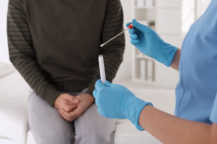
2. During a breast biopsy, after the breast is made numb, a small amount of tissue is removed and looked at under the microscope. This can tell if a lump or suspicious area is cancer or not.
3. An image-guided breast biopsy takes under an hour, requires no preparation, and generally causes little to no discomfort.
At our Breast Center, we’ll do everything we can to make sure you can get your biopsy happens quickly, and that it won’t hurt.
We're Here to Help
Learn about Stamford Health's Breast Center support services.If you have questions, call 203.276.PINK (7465).
Here are few things to keep in mind if you need a breast biopsy.First, the facts:
1. Most women who have breast biopsies DO NOT have breast cancer. In fact, about 4 out of 5 breast biopsies are benign (not cancer).
2. During a breast biopsy, after the breast is made numb, a small amount of tissue is removed and looked at under the microscope. This can tell if a lump or suspicious area is cancer or not.
3. An image-guided breast biopsy takes under an hour, requires no preparation, and generally causes little to no discomfort.
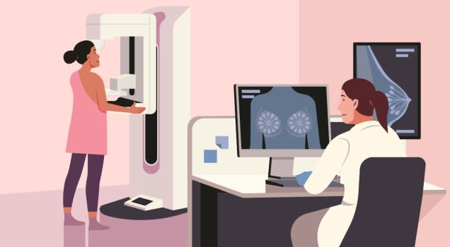
What is a breast biopsy?
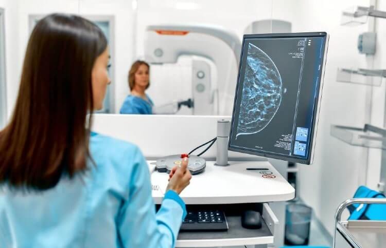
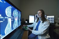
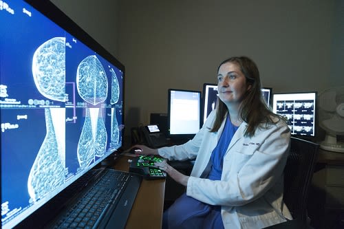
A breast biopsy means that a small piece of tissue is removed from the breast. It is generally done in radiology, using mammography, ultrasound or MRI for guidance. The area is made very numb before a small, hollow needle is inserted into the precise area. This procedure takes much less time than a surgical biopsy, and requires almost no recovery time.Who will perform my breast biopsy?
The Stamford Health Breast Center is unique, in that all image-guided biopsies are performed by fellowship trained radiologists who specialize in, and only do, breast radiology. Each of the radiologists has years of experience performing minimally invasive breast biopsies. Before the procedure, the breast radiologist will discuss the procedure in detail, and answer all of your questions.What happens if I have pain?
Unlike almost anywhere else, our Breast Center has a dedicated nurse that assists in every single breast biopsy. If you have any pain or discomfort at any time, just let the nurse or doctor know, and they will treat you accordingly.My friend had her biopsy using MRI. Why is my biopsy done using Ultrasound?
Each and every patient is unique, and all biopsy procedures are tailored to the individual needs of the patient. In the past, surgeons removed abnormalities through incisions in the breast. Now, when an abnormality is seen by MRI, mammography or ultrasound, a targeted biopsy using a skinny needle, can be precisely placed into the abnormality. If the abnormality is seen best by ultrasound, radiologists use that method to guide the procedure. If it is seen best by MRI or mammography, the biopsy is performed using those imaging tools.Do I need to prepare for a breast biopsy?
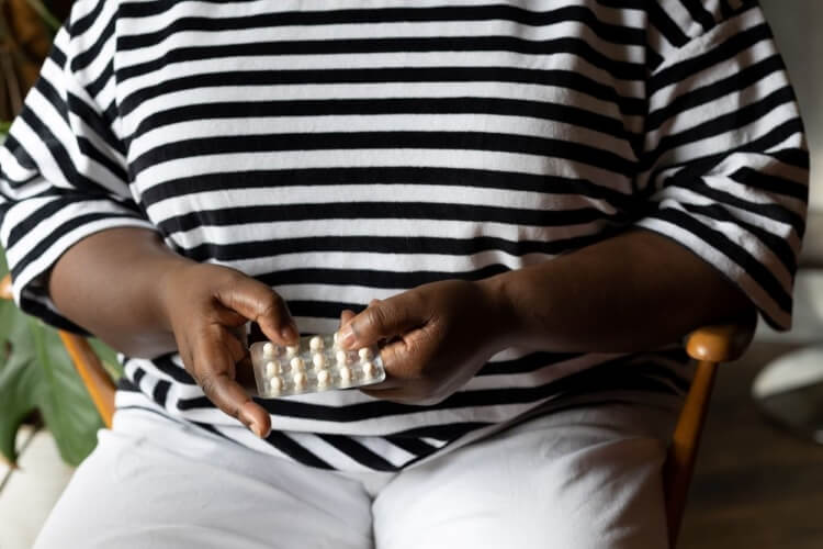
No special preparation is needed for an image-guided breast biopsy. However, when you are scheduling your biopsy, it is very important that you discuss all of the medications you are taking with our team. There are several medications, most commonly Aspirin, Ibuprofin (ie. Motrin, Advil), and other anti-inflammatory drugs; and Coumadin (a blood thinner) that can increase bleeding. In conjunction with your physician, we may ask you to stop your medicine for a few days prior to the biopsy.What will the biopsy be like?
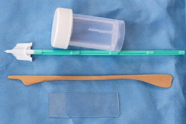
You will be wide awake during the radiology-guided breast biopsy, and should have little or no pain or discomfort. An MRI biopsy and a stereotactic biopsy take less than an hour- and an ultrasound usually takes less than 30 minutes. Before the procedure, we will reimage your breast. We will then clean it well, and make it numb with a very tiny needle. Cells will then be withdrawn through a hollow needle and sent to the pathology lab to examine under the microscope.Why is a marker left behind in the breast?
Following image-guided ultrasound, stereotactic and MRI biopsies, a tiny titanium clip is left behind through the needle at the biopsy site. This is done for two reasons. If the biopsy is benign, we want to be certain we don’t need to biopsy the same spot again. If there are tumor cells (cancer) in the biopsy, the marker is a landmark to guide the breast surgeon to remove the exact area. The clip cannot be felt, does not cause the airport alarms to go off, and is safe for all future tests, including MRI.When will I get results?
After a breast biopsy, the tissue needs to be processed in the laboratory, and then examined under a microscope by one of our highly trained breast pathologists. This can take a few days. As soon as we have the results back from the laboratory, we will call you with the results.How do I know the results are accurate?
At Stamford Health, every single biopsy gets reviewed at a special biopsy meeting- by pathologists, surgeons, nurses and radiologists- to make sure your individual results are accurate. This is a unique benefit of having a biopsy at Stamford- and was cited as a national best practice at a recent program inspection.At our Breast Center, we’ll do everything we can to make sure you can get your biopsy happens quickly, and that it won’t hurt.
Featured Expert/ Author